


Product Description
Endothelial cells lining the microvasculature are known to play a critical “gatekeeper” role in the inflammatory process through their ability to recruit circulating immune cells into tissues and foci of inflammation. Studies show that intestinal microvascular endothelial cells (IMEC) exhibit a strong immune response to LPS challenge and play a critical regulatory role in gut inflammation [1]. Pharmacological inhibition of NOS in activated HIMEC resulted in a significant increase in leukocyte binding [2]. Gene expression profile studies revealed that intestinal endothelial cells express biotinidase, which is involved in biotin recycling [3]. HIMEC cultures have enabled scientists to perform systematic analyses of cytokine profiles with regard to mRNA expression and protein secretion, and to compare these data with cytokine profiles from other endothelial cells.
iXCells Biotechnologies provides high quality HIMEC, which are isolated from human intestinal tissue and cryopreserved at P1, with >0.5 million cells in each vial. HIMEC express vWF/Factor VIII, CD31 (PECAM), and Dil-Ac-LDL by uptake. They are negative for HIV-1, HBV, HCV, mycoplasma, bacteria, yeast, and fungi and can further expand no more than 3 passages in Endothelial Cell Growth Media under the condition suggested by iXCells Biotechnologies. Further expansion may decrease the cell purity.
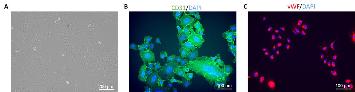
Figure legend: (A) Phase contrast image of Human Intestinal Microvascular Endothelial Cells (HIMEC) at DIV4. (B, C) HIMEC are positive for CD31 (B) and vWF/Factor VIII (C) as shown by immunofluorescence staining.
Product Details
| Tissue | Normal human intestinal tissue |
| Package Size | 0.5 million cells/vial |
| Passage Number | P1 |
| Shipped | Cryopreserved |
| Storage | Liquid nitrogen |
| Growth Properties | Adherent |
| Media | Endothelial Cell Growth Media |
References
[1] Jennifer Y Kasper, Maria Iris Hermanns, Christian Cavelius, Annette Kraegeloh, Thomas Jung, Rolf Danzebrink, Ronald E Unger, Charles James Kirkpatrick. (2016) “The role of the intestinal microvasculature in inflammatory bowel disease: studies with a modified Caco-2 model including endothelial cells resembling the intestinal barrier in vitrohe role of the intestinal microvasculature in inflammatory bowel disease: studies with a modified Caco-2 model including endothelial cells resembling the intestinal barrier in vitro”. Int J Nanomedicine. 11:6353-6364.
[2] David G. Binion, Sidong Fu, Kalathur S. Ramanujam, Yuh Cherng Chai, Raed A. Dweik, Judith A. Drazba, Justin G. Wade, Nicholas P. Ziats, Serpil C. Erzurum, and Keith T. Wilson. (1998) “iNOS expression in human intestinal microvascular endothelial cells inhibits leukocyte adhesion”. Physilogy. 275: G592-G603.
[3] E M Nilsena, Johansen, F L Jahnsen, K E A Lundin, T Scholz, P Brandtzaeg, G Haraldsen. (1998) Cytokine profiles of cultured microvascular endothelial cells from the human intestine. 42: 635-642