
---Mouse%20Brain%20Vascular%20Pericytes.jpg)
Product Description
Brain Vascular Pericytes (BVP) are perivascular cells that are closely associated with the endothelium of capillaries and other small vessels [1]. BVP, located between endothelial cells and astrocytes in the brain, communicate with other cells by extending long cytoplasmic processes which wrap around the capillaries [1, 2]. BVP participate in a variety of processes including angiogenesis, endothelial cell survival, regulation of capillary blood flow, and establishment and maintenance of the blood-brain barrier [3,4]. Pericyte dysregulation has been linked to several pathological conditions such as hypertension, diabetic retinopathy, atherosclerosis, multiple sclerosis, Alzheimer’s disease, and tumor angiogenesis [2, 4]. The unique and diverse functions of BVP make them novel candidates for cell therapy in regenerative medicine. Cultured primary mouse BVP (MBVP) are a useful in vitro model for understanding the molecular mechanisms of blood-brain barrier regulation and for studying a wide variety of central nervous system diseases.
iXCells Biotechnologies provides high quality Mouse Brain Vascular Pericytes (MBVP), which are isolated from adult mouse brain and cryopreserved at P2, with >0.5 million cells in each vial. MBVP express Neural/glial antigen 2 (NG2) and α-smooth muscle actin. They are negative for HIV-1, HBV, HCV, mycoplasma, bacteria, yeast, and fungi and can further expand no more than 3 passages in Mouse Pericyte Growth Medium (Cat# MD-0030) under the condition suggested by iXCells Biotechnologies. Additional expansion may decrease the purity and proliferation rate.
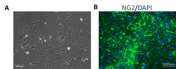
Figure 1. Mouse Brain Vascular Pericytes (MBVP). (A) Phase contrast image of MBVP. (B) Immunofluorescence staining with antibody against NG2.
Product Details
| Tissue | Adult mouse brain |
| Package Size | 0.5 millioncells/vial |
| Passage Number | P2 |
| Shipped | Cryopreserved |
| Storage | Liquid nitrogen |
| Growth Properties | Adherent |
| Media | Pericyte Growth Medium (Cat# MD-0030) |
References
[1] Dore-Duffy P, Cleary K. (2011) “Morphology and properties of pericytes.” Methods Mol Biol. 686:49-68.
[2] Allt G, Lawrenson JG. (2001) “Pericytes: cell biology and pathology.” Cells Tissues Organs. 169: 1-11.
[3] Daneman R, Zhou L, Kebede A, Barres B. (2010) “Pericytes are required for blood-brain barrier integrity during embryogenesis.” Nature. 468:562-566.
[4] Kutcher M, Herman I. (2009) “The pericyte: cellular regulator of microvascular blood flow.” Microvasc Res. 77: 235-246.
Biswas, S., Shahriar, S., Giangreco, N. P., Arvanitis, P., Winkler, M., Tatonetti, N. P., Brunken, W. J., Cutforth, T., & Agalliu, D. (2022). Mural Norrin/β-catenin signaling regulates Lama2 expression to promote neurovascular unit assembly. https://doi.org/10.1101/2022.02.18.481046 -- Learn More
Lee, S. J., Kim, S., Jo, D. H., Cho, C. S., Kim, S. R., Kang, D., Chae, J., Yoo, D. K., Ha, S., Chung, J., & Kim, J. H. (2021). Specific ablation of pdgfrβ-overexpressing pericytes with antibody-drug conjugate potently inhibits pathologic ocular neovascularization in mouse models. Communications Medicine, 1(1). https://doi.org/10.1038/s43856-021-00059-3 -- Learn More
Kaushik, D. K., Bhattacharya, A., Lozinski, B. M., & Wee Yong, V. (2021). Pericytes as mediators of infiltration of macrophages in multiple sclerosis. Journal of Neuroinflammation, 18(1). https://doi.org/10.1186/s12974-021-02358-x -- Learn More
Grant, S., McMillin, M., Frampton, G., Petrescu, A. D., Williams, E., Jaeger, V., . . . DeMorrow, S. (2018). Direct comparison of the Thioacetamide And Azoxymethane models of type A hepatic encephalopathy in mice. Gene Expression, 18(3), 171-185. doi:10.3727/105221618x15287315176503 -- Learn More