

Product Description
Epidermal keratinocyte is the predominant cell type in the outermost layer of the skin – the epidermis, which serves as a critical barrier to separate and protect the inside of human body from outside environment and damage from pathogens, heat, UV radiation and water loss. Epidermal keratinocytes originate in the stratum basale, and they undergo gradual differentiation and migrate towards the surface of the epidermis until they reach the stratum corneum, where they form a tight layer of nucleus-free and highly keratinized squamous cells. This layer forms an effective barrier to prevent water loss and the entry of infectious agents. Keratinocytes are also known to produce various growth factors, cytokines, antimicrobial peptides, and complement factors. Therefore, keratinocytes are important for wound healing, inflammation, infection, skin microbiome and immune response.
iXCells Biotechnologies provides high quality primary Mouse Epidermal Keratinocytes-adult (MEK-a), which are isolated from adult mouse tail and cryopreserved at P0, with ≥0.5 million cells in each vial. MEK-a are characterized by phalloidin staining and are negative for mycoplasma, bacteria, yeast and fungi. MEK-a are not recommended for expanding or long-term cultures.
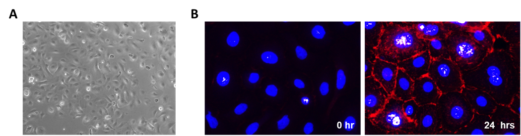
Figure 1. (A). Phase contrast image of primary Mouse Epidermal Keratinocytes – Adult (MEK-a). (B). MEK-a were differentiated in the presence of 0.2mM CaCl2, and cells were stained with phalloidin (red) to visualize the formation of actin fiber-rich filopodial projections between adjacent cells during differentiation. Nuclei were counterstained with DAPI.
Product Details
| Tissue | Epidermis from C57BL/6 mouse tail skin |
| Package Size | 0.5 million cells/vial |
| Passage Number | P0 |
| Shipped | Frozen |
| Storage | Liquid nitrogen |
| Growth Properties | Adherent |
| Media | Keratinocyte Growth Medium (Cat# MD-MD-0047) |
References
[1] Raja, Sivamani K, Garcia MS, Isseroff RR. Front Biosci. 2007;12:2849-68. Wound re-epithelialization: modulating keratinocyte migration in wound healing.
[2] Proksch E1, Brandner JM, Jensen JM. Exp Dermatol. 2008 Dec;17(12):1063-72. The skin: an indispensable barrier.
[3] Pasparakis M, Haase I, Nestle FO. Nat Rev Immunol. 2014 May;14(5):289-301. Mechanisms regulating skin immunity and inflammation.